Wednesday, December 9, 2009

IMAGING OF CEREBRAL HEMORRHAGE AND AV MALFORMATIONS

INTRACEREBRAL HEMORRHAGE
CT Features
br-740.gif
      Together, hypertension, aneurysm, and vascular malformations account for 80% of intracerebral hemorrhages. All cerebral hematomas, whatever the cause, have a similar resolution pattern on CT. The rate of resolution depends on the size of the hematoma, usually within one to six weeks, and they resorb from the outside toward the center. Perihematoma low density appears in 24-48 hours. Rim enhancement appears in one week and persists for six weeks. The end result of a hematoma is decreased parenchymal density, focal atrophy and local ventricular dilatation.

MR Appearance
      Intracerebral hematomas have a very dynamic appearance on MR, changing in signal intensity over time. Acute blood, in the form the oxyhemogloblin, is isointense with the brain parenchyma. Within a few hours, the oxyhemoglobin is converted to deoxyhemoglobin within the hematoma. Deoxyhemoglobin has a predominant effect of shortening T2, resulting in low signal on T2-weighted images. After three to four days, the deoxyhemoglobin is progressively converted to methemoglobin, which is a paramagnetic substance. Although methemoglobin shortens both T1 and T2, the predominant effect is T1 shortening. As a result, at this stage, hematomas are high signal in both T1-and T2-weighted images. Over the next few months, the methemoglobin is slowly broken down into hemichromes which produce only mild T1 shortening. Hematomas at this end stage are slightly high signal on T1-weighted images and remain high signal on the T2-weighted images. Another interesting phenomenon occurs around the periphery of hematomas. Macrophage activity results in degradation of the methemoglobin and conversion of the iron moiety to hemosiderin. Hemosiderin shortens T2 and produces a black ring around the hematoma on T2-weighted images. We have observed this ring as early as nine days after hemorrhage, and the ring becomes thicker over time. The amount of hemosiderin varies from one hematoma to another, and the specific physiologic and chemical factors that influence this are unknown. In small hematomas (less than 1 cm), we have noted low signal intensity from hemosiderin throughout the cavity. The length of time that the hemosiderin will remain in the area of a hematoma is also unknown, but we have observed hemosiderin at the site of a
br-7401.gif

previous hematoma as long as four years following the primary hemorrhage. From this discussion, it is apparent that the specific signal intensities of a hematoma on T1- and T2-weighted images provide a clue as to the age of the hemorrhage. Endnote Endnote

Hypertensive Hemorrhage
      The criteria for hypertensive hemorrhage include a hypertensive patient, 60 years of age or older, and a basal ganglia or thalamic location of the hemorrhage. A CT scan is the procedure of choice for evaluating these patients. Arteriography is necessary only if one of these criteria is missing. Hypertensive hemorrhages are often large and devastating. Since they are deep hemorrhages and near ventricular surfaces, ventricular rupture is common. One-half of hypertensive hemorrhages occur in the putamen; the thalamus in 25%; pons and brainstem, 10%; cerebellum, 10%, and cerebral hemispheres, 5%.

VASCULAR MALFORMATIONS

      Cerebrovascular malformations have been classified into four distinct types including arteriovenous malformation, cavernous angioma, capillary telangiectasia and venous malformation. Capillary telangiectasia is characterized by abnormal, dilated capillaries separated by normal neural tissue. They are usually found in the pons at autopsy and are occult clinically and radiographically. The other three malformations are commonly seen on imaging studies and have distinct features.

br-7402.gif
Arteriovenous Malformation
      The arteriovenous (AV) malformation consists of a congenital abnormality of anomalous, dilated capillaries that result in shunting of blood from the arterial to venous side. AV malformations are by far the most common of the cerebrovascular malfor mations. One-half of patients present with seizures or a neurological deficit due to compression of normal brain or a steal phenomenon. The other half presents with hemorrhage. The hemorrhage is usually more benign than that due to a ruptured aneurysm. Ninety-five percent of AV malformations are in the supratentorial compartment, either in a lobar or deep location and 10% are in the infratentorial region. Dural supply is more commonly found with infra tentorial lesions although it is important to remember than any AV malformation adjacent to a dural surface can receive dural contributions.
      CT features of an AV malformation on plain scan include a high- absorption irregular mass with large feeding arteries and draining veins, focal areas of calcification and no surrounding edema or mass effect. The contrast scan shows serpiginous enhancement with prominent arteries and veins. Due to the rapidly flowing blood from these lesions, a flow void is observed on MR scan. As a result, the characteristic feeding arteries and draining veins can be imaged without any injection of contrast material. Endnote
      The best MRA sequences for depicting the anatomy of AV malformations are 3D TOF and PC methods. The TOF technique may not show the draining veins in their entirety due to saturation effects. Endnote Gd will improve venous visualization, but in general, PC is better for imaging the venous side. By using two different velocity-encoding factors, 80 and 20 cm/sec, arterial and venous phase images can be generated. High flow through these lesions often produce turbulence and some signal loss within the feeding arteries. Selective saturation pulses can be used to isolate arterial supply.
br-7403.gif
      As mentioned earlier, flow voids from AV shunting help identify AV malformations on spin-echo MR scans. In fact, spin-echo imaging is probably more sensitive than MRA for detecting AV malformations. Moreover, MRA probably has a limited role in the initial diagnostic workup of AV malformations because a conventional angiogram is required anyway. The proper role for MRA will likely be to follow these lesions, to assess the affects of radiation therapy or embolization procedures, and to check for growth of partially resected lesions.
      One should suspect AV malformation as a cause of an intracerebral hemorrhage if the hemorrhage is lobar and away from the territory of the anterior communicating and middle cerebral arteries, and also in deep hemorrhages in younger, normotensive patients. It is important to remember that the hematoma may compress a small AV malformation. If the initial angiogram is negative, a follow-up study should be done one to two months later, after the hematoma and mass effect have resolved. AV malformations can thrombose either spontaneously or due to compression by the hematoma.

Cavernous Angioma
br-7404.gif
      They are characterized by a honeycomb of endothelium-lined vascular spaces, separated by fibrous, collagenous bands with no intervening neural tissue. Most cavernous angiomas are asymptomatic and are noted incidentally on MR scans. They may cause seizures or a focal neurologic deficit, and on occasion they will be of sufficient size to produce symptoms by mass effect. The intralesional hemorrhages are usually small and occult clinically. Multiplicity is common.
      Cavernous angiomas invariably contain hemosiderin from chronic hemorrhage and are distinctly hypointense on T2-weighted MR images. Lesion margins are "fuzzy" due to the magnetic susceptibility effect of the hemosiderin, and a "blooming effect" occurs with gradient-echo sequences. Calcification is often present. Mild enhancement can be obscured by the hemosiderin. Endnote
      Larger cavernous angiomas have a more complex appearance from multiple hemorrhages of varying ages. Hemosiderin lines the perimeter of these lesions and also outlines the internal compartments that contain various components of hemorrhage. Endnote

Venous Malformation
      Venous malformations involve only venous structures and are usually incidental findings on MR, CT or arteriography. Venous malformations are less likely to bleed than the arteriovenous variety. Those that are located in the cerebellum or brainstem seem to be at a higher risk for bleeding than the supratentorial ones. Also, cavernous angiomas have been shown to be associated with some venous angiomas, which may increase the risk of hemorrhage. Endnote
      Venous malformations have a characteristic appearance on MR and contrast-enhanced CT scans. A radial pattern of small veins join to form a single large vein that drains toward the cortical surface. Drainage into the deep venous system is uncommon. Usually, flow is sufficiently fast to produce a flow void on spin-echo MR images. They are also routinely visualized with MR angiography. Endnote

HEAD TRAUMA

HEAD TRAUMA

      Patients with head trauma constitute a large percentage of the cases referred for neuroimaging. Initially, the role of MR in these patients was considered limited due to the time required for the examination, difficulty in using life-support and monitoring equipment within the scanning room and problems in imaging acute hemorrhage. While some of these problems still remain, MR has come to be used more frequently in these patients, particularly in the subacute period.
      The most common head injuries result from blunt or non- penetrating trauma. These frequently induce a temporary or longer loss of consciousness and the brain may suffer gross damage despite the lack of a skull fracture or penetrating injury. Skull fractures may serve to indicate the presence of significant trauma but the absence or presence of a skull fracture cannot be used to predict the presence or severity of intracranial injury. Endnote
 

EXTRACEREBRAL HEMORRHAGE

      Acute epidural hematomas are often associated with skull fractures and lacerations of the dural vessels, most often meningeal arteries and veins but occasionally a dural sinus. Two-thirds of epidural hematomas are in the temporo-parietal region and they usually have a biconvex or lentiform configuration. Epidurals are limited by the firmer attachment of the dura at the suture margins, but they may cross the midline, especially with superior sagittal sinus lacerations, and they also can bridge the supra- and infratentorial compartments with tears along the torcula and transverse sinuses.
      Subdural hematomas, both acute and chronic, are most often caused by bleeding from torn bridging dural veins. Subdural hematomas are less frequently associated with skull fractures, but more frequently associated with parenchymal brain damage. The subdural space is a more freely communicating space and the hematomas form a crescentic shaped layer over the brain surface. Subdural hematomas readily cross suture lines but do not cross the midline. Instead, they extend along the dura of the falx into the interhemispheric fissure and onto the tentorium, which epidurals cannot do. Both epidural and subdural hemorrhages occur within the confined space of the bony calvarium and compress the adjacent brain, often requiring emergency evacuation.
      Chronic subdural hematomas are usually related to a slower venous bleed without accompanying cerebral parenchymal injury. A thick,vascular dural membrane forms that can be a source for repeated episodes of hemorrhage. These collections are more often biconvex, rather than the crescentic shape of acute subdural hematomas. The injury leading to a chronic subdural can be relatively minor and may have occurred weeks before presentation. Patients often present with disturbances of mentation and consciousness rather than focal or lateralizing signs. An iatrogenic cause is overshunting or too rapid decompression of chronic hydrocephalus.
      Subdural hygromas or effusions consist of collections of CSF in the subdural spaces, presumably due to a traumatic arachnoid tear or they may also develop following ventricular shunting. They may accumulate slowly during the first few days following head trauma, especially in the pediatric population.
br-400.gif
      Multiple studies have demonstrated improved visualization of extra-axial hemorrhage with MR compared to CT, largely related to the high conspicuity of hyperintense subacute hemorrhage (methemoglobin) on T1-weighted images and the multiplanar capabilities of MR. Coronal images are very helpful for identifying subtemporal collections and hemorrhage adjacent to the tentorium cerebelli. Chronic subdural hematomas are often isointense with gray matter on T1-weighted images, probably due to dilution and partial resorption or breakdown of free methemoglobin. High T1 signal within what otherwise appears to be a chronic subdural hematoma suggests rebleeding. Hemosiderin is rarely seen in subdural hematomas without repeated episodes of bleeding, due to either low macrophage activity or removal of hemosiderin that has formed. The presence of membranous strands coursing through an extra-axial collection is additional evidence for a chronic subdural hematoma. The thick subdural membranes will also enhance following contrast infusion. Endnote
 

BRAIN CONTUSIONS

      Damage to the brain parenchyma is a common component of head trauma. The type, location and degree of brain injury is determined to a large extent by the physical properties of the skull and brain. The skull is very hard and rigid, and protects the brain from direct injury. However, the inner table of the skull has roughened edges and ridges of bone along the floor of the anterior cranial fossa, sphenoid wings and petrous ridges that can contuse the brain surface during the compressive forces of trauma.
      Injury of the brain parenchyma sets in motion a series of events. Tissue disruption and cell injury are associated with release of vasoactive substances and other byproducts. Subsequent increase in vascular permeability to serum proteins results in a progressive increase in interstitial fluid. Over a period of several days, the edema fluid spreads within the white matter, producing mass effect on adjacent structures and possible further damage. More serious injuries may be associated with vascular disruption and hemorrhage into contusions. Cortical contusion are usually multiple, measuring approximately 2-4 cm in size, and 30% to 50% of lesions are hemorrhagic. Approximately 50% to 75% of cortical contusions involve the frontal and temporal lobes, particularly the lateral surfaces of both lobes and the inferior surface of the frontal lobes.
      Varying signal intensity patterns are seen on MR depending on the age and amount of hemorrhage present. In several studies MR has had a decided advantage over CT in the imaging of bland contusions and has been roughly equivalent to CT in imaging hemorrhagic contusions. Overall, MR has shown approximately 90% of all cortical contusions imaged by either modality. In general, T2-weighted images are best for evaluating brain contusions. T1-
br-4001.gif

weighted images are helpful to look for any associated hemorrhage. Nonhemorrhagic contusions are hyperintense on T2 and hypointense on T1-weighted scans due to brain edema and increased water content in the lesions. The brain edema increases during the first few days, producing mass effect on adjacent brain structures. With time, the edema subsides and the dead tissues are removed, resulting in areas of encephalomalacia and compensatory focal dilatation of adjacent ventricles and sulci.
      The MR appearance of hemorrhagic contusions is more dynamic, changing over time as the internal chemistry of the hematoma changes. In fact, the signal intensities on T1 and T2-weighted images often provide clues about the approximate age of hemorrhagic contusions. The central hypointensity of acute hemorrhagic contusions on T2- weighted images is often highlighted by the surrounding edema. After a few days, methemoglobin forms and gives a mottled pattern of high signal on T1-weighted images owing to the multifocal nature of hemorrhage into cortical contusions.
      The brain stem is also subject to injury from head trauma. Although it is protected from direct injury by its location, acceleration/deceleration forces associated with impact to the head may produce displacement and twisting of the brain stem. These forces can result in tearing of penetrating arteries or veins, and compression of the brain stem against the sharp edges of the tentorium or surfaces of the clivus and petrous bones.
 

SHEAR INJURIES

      Severe head injuries are often associated with rotational forces that produce shear stresses on the brain parenchyma. The brain itself has very little rigidity and is extremely incompressible. Brain volume can be decreased only by exerting great pressure. On the other hand, the brain is soft and malleable. Relatively little effort is required to distort the shape of the brain. The parenchyma is of relatively uniform density, except for differences between the CSF of the ventricles and surrounding brain tissue. Slight differences in density also exist between gray and white matter.
      When the skull is rapidly rotated, it carries along the superficial brain parenchyma but the deeper structures lag behind, causing axial stretching, separation and disruption of nerve fiber tracts. Shear stresses are most marked at junctions between tissues of differing densities. As a result, shear injuries commonly occur at gray/white matter junctions, but they are also found in the deeper white matter of the corpus callosum, centrum semiovale, brain stem (mostly the midbrain and rostral pons) and cerebellum. Lesions in the basal ganglionic regions are usually found along the borders between the ganglia and the internal or external capsules, in other words, the deep gray-white matter junctions of the cerebral hemispheres. The thalamic and basal ganglia injuries are hemorrhagic in slightly more than 50% of cases. On the other hand, shear injuries of the corpus callosum and centrum semiovale are more often nonhemorrhagic.       Attempts to correlate CT findings with acute and chronic sequelae of closed head trauma have been discouraging, largely related to the insensitivity of CT to many cerebral injuries. Chiefly among these, poorly seen by CT and well seen by MR, are the diffuse axonal injuries or white matter shear injuries. These injuries constitute the most frequent findings on MR in head trauma, comprising as high as 40% of all lesions. Shear injuries are most often multiple, ovoid and parallel to white matter fiber bundles. They are hyperintense on T2 and hypointense of T1-weighted scans, unless hemorrhagic components are present, in which case more complex patterns are observed. During transition phases of hematoma evolution, combinations of methemoglobin, hemosiderin rings and peripheral edema can result in layers of differing signal intensity and a target-like appearance. The axial plane is the primary plane of imaging for both cortical contusions and shear injuries, but supplemental coronal views are helpful to assess injuries to the body of the corpus callosum and the inferior frontal and temporal lobes. Fast scan techniques or gradient-echo images have lower resolution but are useful in uncooperative patients. Contrast enhancement has little role in the evaluation of brain contusions. Endnote
 

CHILD ABUSE (Non-accidental trauma)

      Unfortunately, there are over a million cases of child abuse in the United States each year. Intracranial injuries may be the result of either direct blows to the head, shaking, whiplash, or strangulation. The presence of an inner hemispheric subdural hematoma is often a tip-off of underlying abuse as it results usually from a whiplash type of injury rather than from a direct blow. There may also be associated subarachnoid hemorrhage as well as subdural hematomas or even subdural hygromas of different ages. One should also be alert for possible underlying non-accidental trauma in the presence of multiple fractures often of different ages. Cerebral anoxia and/or infarction (reversal sign) can occur as a result of strangulation. One should be on the alert for other sites of injury throughout the body.
 

PENETRATING TRAUMA

      The impact cavity of a gunshot wound to the head may vary with the shock wave, the bullet and/or bone fragments. One should try to identify the site of injury, the number of fragments, the location, the path of the wound, and the nature of the hemorrhage. One should also be alert to the path of injury relative to dural sinuses as venous infarctions and/or epidural hematomas may result as well as paranasal sinuses as this can be a source for infections. One should remember that gun shot wounds to the head are not necessarily always fatal.
 

VASCULAR INJURIES

      Traumatic carotid cavernous fistulas can result from an injury to the carotid artery at the level of the cavernous sinus. This causes a high flow arterial venous shunt and often results in painful unilateral pulsating exophthalmos as a result of increased flow in a retrograde direction in the superior ophthalmic vein. This can lead to visual loss. Alternatively, the arterial venous shunt can extend into the inferior petrosal sinus and then into cortical veins of the cerebellum which can lead to subsequent hemorrhage.
      Traumatic dissections of the carotid or vertebral arteries in the neck can cause stroke. This can be due to strangulation or whiplash mechanisms which may tear the intima. Typically the carotid is dissected usually just proximal to entering the petrous bone at about the C1-C2 level.
      Post-traumatic pseudoaneurysms can occur as a result of vascular injuries intracranially. These do not have to be located at more typical locations for nontraumatic aneurysms around the circle of Willis. Vascular spasm can occur as a result of subarachnoid hemorrhage. Endnote

SPECIAL MR TECHNIQUES

      Diffusion weighted images (DWI) help elucidate the complex nature of brain injury. First, several events associated with trauma can result in ischemic brain injury, including cardiac arrest, hypotension, hypovolemia, hypoxia/anoxia, major vascular laceration, distal embolus, fat emboli, penetrating vascular injury, and herniation syndromes causing vascular compromise. Ischemia appears as high signal on DWI and hypointensity on apparent diffusion coefficient (ADC) maps. Non-ischemic traumatic lesions (contusions, DAI) have variable diffusion properties. If the cells and axons are completely disrupted, leakage of intracellular fluid into the extracellular space results in vasogenic edema and high signal intensity on both DWI and ADC images. On the other hand, minor mechanical damage may stun cell metabolism and the cell membrane fluid transport system, leading to influx of sodium and cytotoxic edema. Finally, microvascular arterial injury may produce an element of true ischemia, whereas microvascular venous injury would lead to vasogenic edema. Endnote , Endnote

      Diffusion tensor imaging (DTI) has demonstrated high sensitivity for axonal disruption and is especially helpful for detecting subtle injuries. MR spectroscopy can assess the overall disease burden in traumatic brain injury. Measures of brain NAA levels have been shown to correlate with neuropsychological function.


IMAGING OF STROKE AND CEREBRAL ISCHEMIA

 The importance of occlusive cerebral vascular disease is confirmed by two facts: 1) stroke is the third commonest cause of death in the United States; and 2) one-half of neurology inpatients have stroke related problems. These patients present with either asymptomatic bruits, transient ischemic attacks (TIA), or stroke. A TIA is a transient neurologic deficit lasting from a few seconds to a few hours. Another terminology that is sometimes used is reversible ischemic neurologic deficit (RIND), which refers to a neurological deficit that lasts longer than 24 hours, but results in complete recovery. These patients are often included in the larger category of stroke. The neurological deficit may consist of visual disturbances (transient monocular blindness or amaurosis fujax), weakness or numbness in an extremity or aphasia. Strokes in a carotid territory usually result in a contralateral hemiparesis and, if in the dominant hemisphere, an aphasia. Vertebral-basilar ischemia results in dizziness, diplopia, dysarthria and weakness or numbness that does not lateralize well.

CAUSES OF STROKE

      The five major causes of cerebral infarction are vascular thrombosis, cerebral embolism, hypotension, hypertensive hemorrhage, and anoxia/hypoxia. Thrombotic strokes may occur abruptly but the clinical picture often shows gradual worsening over the first few hours. Primary causes of arterial thrombosis include atherosclerosis, hypercoagulable states, arteritis, and dissection. Secondary compromise of vascular structures can result from traumatic injury, intracranial mass effect, neoplastic encasement, meningeal processes, and vasospasm.
      Embolic strokes characteristically have a very abrupt onset. After a number of hours, there may be sudden improvement in symptoms as the embolus lyses and travels more distally. The source of the embolus is usually either the heart (patients with atrial fibrillation or previous myocardial infarction) or ulcerated plaques at the carotid bifurcation in the neck.
      Hypotension can be cardiac in origin or result from blood volume loss or septic shock. Hypertension can cause a primary intracerebral hemorrhage, or the elevated arterial pressure can overwhelm the brain's autoregulatory mechanism, resulting in breakthrough of the blood-brain barrier and brain edema. The latter phenomenon of hypertensive encephalopathy is a potential complication of eclampsia, but is usually transient and reversible. Anoxia/hypoxia events are usually related to respiratory compromise from severe lung disease, perinatal problems, near drowning, high altitude, carbon monoxide inhalation, or CNS mediated effects.

PHYSIOLOGY, METABOLISM, AND PATHOLOGY

      The brain requires glucose and oxygen to maintain neuronal metabolism and function. Hypoxia refers to inadequate delivery of oxygen to the brain, and ischemia results from insufficient cerebral blood flow (CBF). Normal CBF is 50-55 ml/100gm/min. If the CBF drops below 18, electrical activity ceases. If the CBF dips below 10, neuronal metabolism stops.
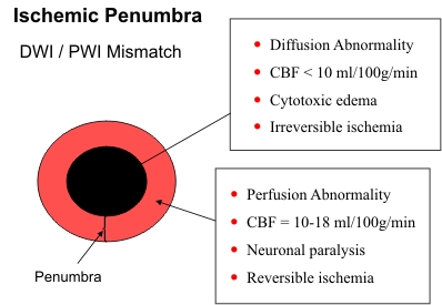
      The consequences of cerebral ischemia depend on the degree and duration of reduced CBF. Neurons can tolerate ischemia for 30-60 minutes. Perfusion must be reestablished before 3-6 hours of ischemia have elapsed or before the CBF drops to 10. The range of CBF between 10 and 18 has been called the "ischemic penumbra" because the neuronal damage is mild and reversible if flow is restored within a few hours. Clinically, a window of opportunity is available to intervene therapeutic and to prevent the ischemic brain tissue from going on to infarction.
      If flow is not reestablished to the ischemic area, a series of metabolic processes ensue. The neurons become depleted of ATP and switch over to anaerobic glycolysis, a much less efficient pathway. Lactate accumulates and the intracellular pH decreases. Without an adequate supply of ATP, the membrane ion pump fails. There is an influx of sodium, water, and calcium into the cell. The excess calcium is detrimental to cell function and contributes to membrane lysis. Cessation of mitochondrial function signals neuronal death. The astrocytes and oligodendroglia are slightly more resistant to ischemia, but their demise follows shortly if blood flow is not restored.
      Pathologic changes within the neuropil follow the metabolic abnormalities. One of the first effects is cytotoxic edema that results from failure of the Na/K ion pump. Early on, this stage is still reversible. Prolonged ischemia leads to cell death and coagulation necrosis. After 3-6 hours of ischemia, irreversible damage occurs to the capillary endothelium. The blood-brain barrier becomes dysfunctional and serum proteins and water leak into the interstitial space. Some reperfusion is required to produce vasogenic edema. In fact, vasogenic edema is maximal when residual CBF is between 5 and 10. There is also an influx of macrophages to clean up the dead tissues. Capillary proliferation begins near the end of the first week. The end result of cerebral infarction is an area of encephalomalacia with some surrounding gliosis. The amount of gliosis depends on the number of surviving astrocytes. Endnote
br-710.gif

CT AND MR IMAGING
Acute Infarcts
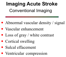
     CT and MR scans in patients with asymptomatic bruits or TIA's are usually negative, unless they disclose abnormalities related to previous events. In patients with stroke, the earliest sign may be abnormal vascular density/signal. Acute thrombus or embolus is hyperdense on CT. Acute clot may be difficult to detect on MR, but the occluded artery should be apparent by the absence of a normal flow void. The absent flow void is easiest to see in the larger arteries at the base of the brain on T2-weighted images. It is not possible to conclusively distinguish a complete occlusion from a critical stenosis with markedly reduced flow. Subacute clot is hyperintense and is easiest to visualize in the basilar and middle cerebral arteries on T1-weighted images. One must be careful not to mistake in-flow enhancement with intraluminal clot. This phenomenon is most often observed in the end slices of a multislice set in arteries with slow flow entering the imaging volume.
      Another valuable sign of acute stroke is arterial enhancement. With slow arterial flow, the spin-echo is able to capture the intravascular signal, and the T1 shortening effect of the gadolinium renders the arteries hyperintense on T1-weighted images. Arterial enhancement is more apparent in the smaller distal branches. It will be present in up to 45% of patients during the first week. Endnote
      The first parenchymal changes observed on CT and MR reflect the cytotoxic edema affecting primarily the gray matter. It is important to remember that the CT scan may be negative for the first 24-36 hours. Massive infarctions may be visible as early as 6 hours. The MR scan is usually positive within three to four hours following a stroke. One of the earlier signs on CT is loss of the normal gray-white contrast as the edematous cortex becomes isodense to the underlying white matter. A similar phenomenon is not observed on MR because the increased water in the gray matter renders the cortex higher signal on T2-weighted images and lower signal on T1-weighted images, thereby increasing gray-white contrast. It is often easier to appreciate the increased cortical signal on proton density-weighted images. The cortical swelling is more apparent on T1-weighted scans. Cortical edema produces effacement of the sulci on both CT and MR. Endnote
      After 6-8 hours the accompanying vasogenic edema highlights the areas of brain infarction. These fluid shifts are more profound and are responsible for effacement of the ventricles and midline shifts. The mass effect increases over the first few days and becomes maximal at about five days.
      Incomplete ischemia has received special attention in the neuroradiologic literature. It results from transient interruption of the blood supply from iatrogenic causes or from an embolus that breaks up and rapidly restores perfusion to the area. The distinctive features of incomplete ischemia are early intense parenchymal enhancement and little or no arterial enhancement. If arterial enhancement is present, it disappears within 24-48 hours. The parenchymal enhancement results from a combination of reperfusion and vasomotor deregulation or vasodilatation. This "luxury of perfusion" last about 24 hours. There may be some mild brain edema during the first 24-48 hours, but the parenchymal signal changes are minimal or absent. Incomplete ischemia is associated with a good prognosis. Endnote

Subacute and Chronic Infarcts
      The subacute stage begins during the second week with capillary proliferation in the area of infarcted brain tissue. This neovascularity is devoid of any blood-brain barrier and intravascular contrast freely diffuses into the interstitial spaces. The serpiginous character of the gyral enhancement is quite distinctive of cerebral infarction. A focal cerebritis or encephalitis can mimic this pattern, but usually the clinical picture sets apart these entities. Following contrast infusion, infarcts will typically enhanced between 2 and 8 weeks, but the enhancement can persist for up to three months.
      As an infarct evolves, it becomes progressively lower in density on CT (higher in signal on T2-weighted images) and more well defined over the next few weeks, eventually approaching the density of CSF. As the mass effect resolves and the infarcted tissue is resorbed, the adjacent sulci and ventricle will enlarge. The end result is a chronic infarct with focal areas of cystic encephalomalacia and some surrounding parenchymal change due to gliosis.

Vascular Patterns
      Since most infarcts result from occlusion of vessels, the CT or MR pattern of abnormality should follow one of the major vascular territories, such as the anterior cerebral, middle cerebral or posterior cerebral arteries. Infarcts can usually be distinguished from inflammatory and neoplastic disease because unlike the white matter pattern of edema found with tumors and abscesses, infarcts involve the cortex as well and, therefore, the abnormal density or signal intensity should extend peripherally to involve the cortex. As mentioned above, the enhancement pattern of infarcts is also fairly characteristic, having a gyral pattern of enhancement along the cortex. If a stroke is due to systemic hypotension or hypoxia, the area of infarction is commonly found in watershed areas between the major vascular territories.
     Lacunar infarction results from occlusion of the small penetrating arteries at the base of the brain, including the lenticulostriate and thalamoperforating arteries. They are smaller infarcts (less than 1 cm) and are found in the basal ganglia, thalamus and brainstem. MR is far more sensitive than CT for detecting small lacunar infarcts, particularly in the brainstem where CT scans are often degraded by artifacts from the bone at the skull base. Endnote

Hemorrhagic Stroke
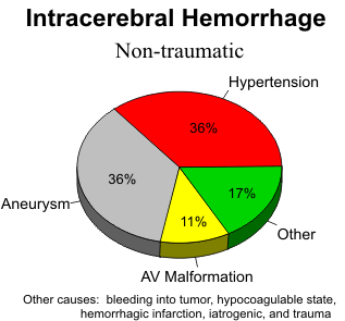
      The four major causes of hemorrhagic stroke are hypertension, hemorrhagic infarction, hypocoagulable state, and amyloid angiopathy. The criteria for hypertensive hemorrhage include a hypertensive patient, 60 years of age or older, and a basal ganglia or thalamic location of the hemorrhage. A CT scan is the procedure of choice for evaluating these patients. Arteriography is necessary only if one of these criteria is missing. Hypertensive hemorrhages are often large and devastating. Since they are deep hemorrhages and near ventricular surfaces, ventricular rupture is common. One-half of hypertensive hemorrhages occur in the putamen; the thalamus in 25%; pons and brainstem, 10%; cerebellum, 10%, and cerebral hemispheres, 5%.
      In stroke patients, despite the fact that the CT is often negative for the first 24-48 hours, it is often obtained on the day of admission to exclude an intracerebral hemorrhage before the patient is placed on anticoagulant therapy. Hemorrhage into an infarct can occur during the first week, usually between the third and fifth days. Hemorrhagic infarction is a hallmark of embolic infarction. This occurs after the embolus breaks up, resulting in reperfusion of the infarcted area. As mentioned above, hemorrhage is also common with venous infarction. Endnote
      Amyloid angiopathy occurs in the elderly, generally over 70 years of age, and is associated with Alzheimer's disease. It results from deposition of eosinophilic material in the media and adventitia of small arteries and arterioles of the cortex and leptomeninges. Fibrinoid degeneration and microaneurysmal dilatation lead to vessel rupture and hemorrhage. The typical imaging pattern is multiple, usually relatively small, cortical hemorrhages that spare the white matter and cerebellum.

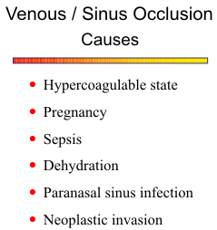
Venous and Dural Sinus Thrombus
br-7101.gif
       Occlusion of the venous sinuses results in cerebral venous engorgement, brain swelling, and increased intracranial pressure. If the thrombosis extends retrograde and involves the cortical veins, secondary cerebral infarction can occur.
      Acute thrombus is hyperdense on CT and may be detected within one of the major sinuses or cortical veins. The other classic sign is the "empty delta" sign due to nonfilling of the superior sagittal sinus on a contrast scan. Nonetheless, MR is far superior for diagnosing abnormalities of the cerebral veins and sinuses. Normally, the dural sinuses have sufficient flow to exhibit a flow void. If that flow void is missing or if the sinuses are hyperintense, thrombosis should be suspected. One must be careful to exclude the possibility of any in-flow enhancement effect. The diagnosis must be confirmed with gradient-echo techniques or MR angiography. Phase-contrast MRA is the preferred technique because it is not adversely affected by intraluminal clot. Endnote
      Associated parenchymal infarcts are found in the areas of venous abnormalities, and the infarcts are often hemorrhagic because arterial perfusion is maintained to the damaged tissue. In cases of superior sagittal sinus thrombosis, the infarcts are typically bilateral and in a parasagittal location.

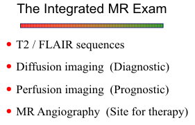
DIFFUSION & PERFUSION IMAGING

      Conventional CT and MR imaging are not sufficiently sensitive to evaluate acute stroke. CT is perfectly adequate to detect intracranial hemorrhage, but in the case of nonhemorrhagic stroke, the CT scan may be negative for the first 24 to 36 hours. FLAIR and T2-weighted images can detect acute stroke by 6 to 12 hours, but most new stroke therapies focus on the first 3 hours after onset. The ultimate goal for imaging is to define the area of brain infarction and perfusion deficit, and to identify any ischemic tissue that can be salvaged by medical or surgical therapy. Diffusion-weighted imaging can detect acute brain infarction within 1 to 2 hours. Perfusion imaging is positive immediately following an acute stroke.

      Diffusion-Weighted Imaging
      Diffusion imaging makes use of the variability of “Brownian motion” of water molecules in brain tissue. Brownian motion refers to the random movement of molecules. Water molecules are in constant motion, and the rate of movement or diffusion depends on the kinetic energy of the molecules and is temperature dependent. In biological tissues, diffusion is not truly random because tissue has structure. Cell membranes, vascular structures, and axon cylinders, for example, limit or restrict the amount of diffusion. Also, chemical interactions of water and macromolecules affect diffusion properties. Therefore, in the brain, water diffusion is referred to as “apparent diffusion.”
      To obtain diffusion-weighted images, a pair of strong gradient pulses are added to the pulse sequence. The first pulse dephases the spins, and the second pulse rephases the spins if no net movement occurs. If net movement of spins occurs between the gradient pulses, signal attenuation occurs. The degree of attenuation depends on the magnitude of molecular translation and diffusion weighting. The amount of diffusion weighting is determined by the strength of the diffusion gradients, the duration of the gradients, and the time between the gradient pulses.
      Diffusion imaging is performed optimally on a high-field (1.5 T) echo-planar system, but it can be accomplished with a turboSTEAM sequence on systems with conventional gradients.
      The diffusion data can be presented as signal intensity or as an image map of the apparent diffusion coefficient (ADC). Calculation of the ADC requires 2 or more acquisitions with different diffusion weightings. A low ADC corresponds to high signal intensity (restricted diffusion), and a high ADC to low signal intensity on diffusion-weighted images.
      In the setting of acute cerebral ischemia, if the cerebral blood flow is lowered to 10 ml/100gm/min, the cell membrane ion pump fails and excess sodium enters the cell, which is followed by a net movement of water from the extracellular to intracellular compartment and cytotoxic edema. Diffusion of the intracellular water molecules is restricted by the cell membranes. The restricted diffusion results in a decreased ADC and increased signal intensity on diffusion-weighted images. Severe ischemia can lower the ADC by as much as 56% of normal tissue at 6 hours. Endnote
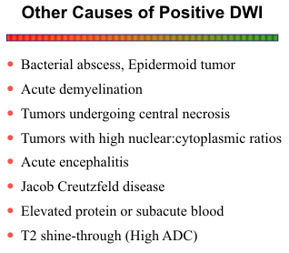
      In patients who present with symptoms of cerebral ischemia, diffusion-weighted images are very helpful to identify any area of acute ischemia and to separate the acute infarction from old strokes and other chronic changes in the brain. Only the acute infarcts appear hyperintense on the diffusion
br-7102.gif

images. Subacute and chronic infarcts, vasogenic edema, the punctate and confluent changes of deep white matter ischemia, and dilated VR spaces are not bright. Endnote Bacterial abscesses may exhibit restricted diffusion due to thick cellular debri within the central cavity. Other diseases of the brain, such as non-bacterial infections, neoplasia, contusions, and demyelinating diseases, are not associated with cytotoxic edema, and therefore as a rule, they are not hyperintense on the diffusion images. One exception is epidermoid tumors, which have restricted diffusion due to the waxy consistency of their contents. Also, the central portions of some primary and secondary brain tumors may exhibit restriction diffusion as they outgrow their blood supply and become ischemic. Occasionally, an acute MS plaque may be mildly hyperintense with diffusion weighting.
      Lesions with prolonged T2 relaxation times are commonly mildly hyperintense on diffusion-weighted images. This phenomenon of “T2 shine-through” can easily be distinguished from true restricted diffusion on the ADC map. Only true restricted diffusion is low signal on the ADC map.

      Perfusion Imaging
      Measurements of brain perfusion include vascular transit time, cerebral blood volume, and cerebral blood flow. The ultimate goal is absolute cerebral blood flow, but it requires serial analysis of arterial input. Most methods measure relative blood flow and compare the two hemispheres or the individual lobes of the brain for regional differences. Both PET and Xenon CT are good techniques, but neither is widely used. PET requires access to a cyclotron and injection of a radioisotope. Xenon CT can be done on a conventional scanner with some additional hardware and software, but use of the xenon gas is cumbersome and the gas must be disposed of properly.
      Vascular transit time and cerebral blood volume are less elegant than cerebral blood flow, but they are useful measurements and can be obtained easily on conventional CT and MR systems with a single bolus injection of contrast material. For CT 40 ml of contrast (300-370 ml I/ml) is injected at a rate of 5-8 ml/sec. Depending on the scanner configuration and number of detector rows, 2-4 sections, 5-10 mm thick are obtained. One image set is acquired per second for 40 seconds. As the bolus of contrast material passes through the cerebral circulation, a transient increase in attenuation or density occurs. A time-density curve is generated for each voxel in each CT slice (see diagram). From the curves, several perfusion image maps can be produced. The time-to-peak (TTP) is the time from the start of the scan until maximum attenuation occurs. The mean transit time (MTT) is the time it takes the contrast bolus to pass from the arterial to the venous side of the cerebral circulation. Assuming a symmetrical attenuation curve, the MTT is measured from the first detection of contrast to the maximum attenuation. The entire area under the curve is a measure of relative cerebral blood volume (rCBV). Finally, a measure of relative cerebral blood flow (rCBF) is calculated by dividing the rCBV by the MTT.
br-7103.gif
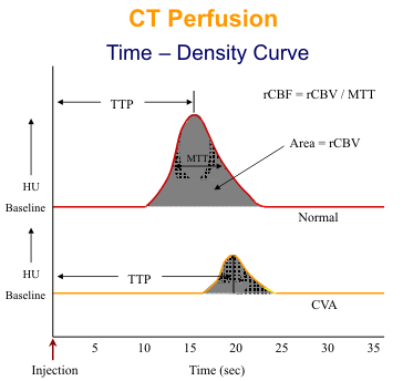
      For MR perfusion, 20 ml of gadolinium is injected at 4ml/sec. An echoplanar sequence is used to acquire 10- 15 slices every 2-3 seconds for 40 second acquisition time. As the paramagnetic gadolinium bolus passes through the cerebral circulation, signal attention occurs due to T2* effects. The time- intensity curves for EPI perfusion are similar to the time-density curves for CT except that the curves are inverted.
      TTP and MTT are very sensitive to hemodynamic changes in the cerebral circulation. Mild increases of these parameters with normal rCBF and rCBV likely indicate that no brain tissue is at immediate risk of infarction. When progresses arterial obstruction, the rCBF decreases followed by the rCBV. The rCBV initially remains normal or even increases due to autoregulation and dilatation of the capillary bed. If both rCBF and rCBV are reduced, tissue must be assumed. If both parameters are markedly reduced, irreversible ischemia has likely occurred.
      Another MR perfusion method is called EPI signal targeting with alternating radiofrequency (EPISTAR). In-flowing blood is tagged with a 180 degree pulse during alternate acquisitions through the imaging plane of interest. The tagged and untagged images are subtracted to obtain the blood volume map. Endnote

      Diffusion/Perfusion Mismatch
br-7104.gif
The difference between the diffusion and perfusion abnormalities provide a measure of the ischemic penumbra or the brain tissue at risk for infarction (see diagram). Restricted diffusion generally indicates unsalvageable brain tissue that is destined for infarction. The perfusion abnormality encompasses all regions with reduced cerebral blood flow. If the diffusion abnormality matches the area of decreased perfusion, no salvageable ischemic brain tissue is present. If the perfusion abnormality is larger than the area of restricted diffusion, the difference identifies the region of reversible ischemia that can be saved if blood flow is re-established promptly. Endnote


VASCULAR IMAGING

     The approach to imaging the cerebral circulation varies from one institution to another. It depends on regional philosophies of therapy in occlusive vascular disease and on the relative strengths of the different imaging procedures at the institution. In any case, one needs to have a rational approach and I will present you with one such approach. If a reliable noninvasive laboratory is available, preferably with duplex Doppler capability, asymptomatic bruits should be evaluated in the noninvasive laboratory. If these results are positive or equivocal, then one should proceed to an angiographic study, either MR or CT angiography. As the technology advances, MRA and CTA are assuming more prominent roles in evaluating occlusive vascular disease. If a patient presents with a stroke, a similar algorithm can be followed. The controversy arises in the patients who present with TIA's. If a patient has had a single TIA, I think it is reasonable to follow these patients in the noninvasive laboratory, but MRA or CTA are other alternatives. With multiple TIA's, an angiographic study should be done. Since the NASCET (North American Symptomatic Carotid Endarterectomy Trial) study Endnote has proved the efficacy of carotid endarterectomy for stenoses ≥70%, accurate assessment of the carotid artery is of paramount importance. MRA and CTA are entirely adequate for evaluating the carotid bifurcations and the circle of Willis. Conventional catheter angiography is generally reserved for cases where some intervention is being planned, such as thrombolysis, clot removal, or angioplasty. Conventional angiography is still required in cases of suspected cerebral arteritis where the more peripheral arteries need to be examined.

MR Angiography
      The two primary methods for MRA are time- of-flight (TOF) and phase contrast (PC). Both use GRE pulse sequences and either 2D or 3D volume acquisitions. TOF makes use of the flow enhancement phenomenon to produce the vascular contrast. PC employs bipolar gradient pulses to tag moving protons with phase shifts. Both produce the MR angiogram by subjecting the data sets to a maximum intensity projection (MIP) ray tracing algorithm to display the vessels from multiple angled views around an axis.
      Imaging the neck vessels takes about 10 minutes and can be done in conjunction with a spin-echo scan of the brain. 2D TOF is very good for imaging this area. Overall, accuracy is quite good for estimating stenoses, although the method tends to overestimate the degree and length of stenosis. Very slow flow distal to a critical stenosis is difficult to detect and may be confused with a complete occlusion.
br-7105.gif
      Gadolinium-enhanced MRA can be obtained with a large field of view to visualize the carotid and vertebral arteries from the aortic arch to the skull base. A 20 cc bolus injection of gadolinium is followed by a rapid 3D acquisition during passage of the contrast through the arch and neck arteries. The high contrast-to-noise images are very good for screening for cerebrovascular disease. Tight stenoses, slow flow, and areas of flow stasis are more accurately assessed with this technique. Endnote
      Another application of MRA is in cases of suspected dissection. Relatively minor trauma is sufficient to cause a dissection, or it can be spontaneous. The MRA may demonstrate complete occlusion or only narrowing of the arterial lumen. Spin-echo images should also be obtained because they are very sensitive for detecting the intramural hemorrhage. The typical appearance of an crescent-shaped hyperintensity with an eccentrically placed flow void may be more convincing for a dissection than the MRA. The MRA is very useful for following a dissection to look for recanalization of a complete occlusion or resolution of the vascular compromise caused by the intramural thrombus.
      MRA can also evaluate the major intracranial arteries about the circle of Willis. The resolution is lower, and flow artifacts limit accurate assessment of arteriosclerotic disease. Nevertheless, MRA is a very acceptable method for imaging the vertebrobasilar system, which is inaccessible to ultrasound and has no effective surgical therapy. The phase images of 2D PC MRA can be used to determine direction of collateral flow about the circle of Willis.
      TOF are PC MRA are very effective in evaluating intracranial veno-occlusive disease, such as superior sagittal sinus thrombosis. PC methods with lower velocity encoding (VENC) factors (15-20 cm/sec) work well for this disease and avoid any possible confusion with thrombus. Thin slab 2D PC can image the full extent of the sagittal sinus in only a few minutes.

CT Angiography
      CT angiography is an alternative to MRA. Following a bolus injection of 80-100 ml of iodinated contrast at 3-4 ml/min, a helical acquisition is performed from the lower neck up to the circle of Willis. Images are reconstructed with a 1.25 mm slice thickness at 0.6-1.0 mm increments. In addition to these source images, sagittal reformatted images are obtained to profile the carotid bifurcations. 3D rendered images are also helpful, but they require a tedious interactive approach to carve off the calcified plaques and bone at the skull base.
      In general, contrast MRA is a more robust technique for evaluating the carotid arteries. The MIP reconstructions are automatically done by the imaging software. No additional physician time is required for off-line image processing. The entire vascular tree from the aortic arch to the circle of Willis is imaged with a single data acquisition. CTA gives a very good view of the carotid bifurcation and the arteries above the skull base.

REFERENCES

Wednesday, October 7, 2009

BOSNIAC CLASSIFICATION OF RENAL CYSTS

Renal cysts are very common, occurring in approximately 1/3 to ½ of adults over 50 years of age. The cysts are typically classified according the Bosniak criteria, as follows: 
Class I cysts have a thin or imperceptible wall, no septations, and is of fluid density. 
Class II cysts still demonstrate no enhancement, but may have thin septa and high density (60 to 90 HU). They may demonstrate calcification or minimal irregularity. 
Class III cysts have increased irregularity, thick septa with or without enhancement, or manifest as a nonenhancing multilocular mass. 
Class IV cysts are malignant lesions with irregularity and enhancement and necrotic/cystic components. 
Various levels of further evaluation are required for Classes II-IV, but no followup for simple cysts is recommended, as their benignity is almost certain. Simple cysts are thought to arise from benign exuberant proliferation of tubular epithelium along with excessive fluid excretion. These lesions can grow to be quite large (several centimeters) causing mass effect, or become infected, but in the vast majority of cases will cause no physical symptoms.
Radiologic overview of the diagnosis:
Cysts can manifest on a variety of modalities, including nephrotomography, nuclear medicine renal scans, MR, CT, ultrasound and even occasionally by plain film. IVP findings can include the "beak" or "claw" sign of parenchymal deformity at the edges of the cyst. CT is the best imaging modality for diagnosis, with a sensitivity nearing 100%. Noncontrasted and 90 second delay post-contrast images are recommended for accurate CT characterization. Ultrasound sensitivity is likely as high as 98% and can often better define the internal characteristics of cysts, but this study is more limited by patient body size.
Key points:

  • Simple cysts are very prevalent in older adults (up to 50% in those older than 50).
  • Cysts can manifest on a variety of imaging modalities, including IVP.
  • CT and ultrasound are best for diagnosis and accurate characterization.
  • Bosniak classification of Classes I-IV are used to characterize the level of likelihood of cyst malignancy.

Wednesday, September 2, 2009

SIGNS IN MUSCULO SKELETAL DISEASES

Alphabetic Listing
Absent Bow Tie Sign     Bucket-handle tear of the menisci
Anteater Nose               Calcaneonavicular coalition
Anterior Drawer Sign     ACL Tear
Bamboo Spine               Ankylosing Spondylitis
Bite Sign                        Avascular Necrosis
Blade of Grass Sign        Paget's Disease
Blister of Bone               Aneurysmal bone cyst
Bone Bruise Sign           ACL Tear
Bone in Bone Sign        Osteopetrosis
Boutonniere Deformity  Rheumatoid arthritis
Bow Tie Sign                Discoid Meniscus
Brim Sign                      Paget's Disease
Button Sequestrum        Eosinophilic granuloma
C Sign                          Subtalar coalition
Cockade Image            Intraosseous lipoma
Cortical Ring Sign         Rotary subluxation
Cotton Wool Sign        Paget's Disease
Crescent Sign              Avascular Necrosis
Crowded Carpal Sign                   Volar Perilunar dislocation
Cupid's Bow Contour                   Normal spine
Dagger Sign                Ankylosing Spondylitis
Deep lateral femoral notch sign       ACL Tear
Double PCL Sign         Bucket-handle tear of the menisci
Drooping Shoulder Sign                 Inferior subluxation of the shoulder
Elbow Fat Pad Sign                       Elbow effusion
Fallen Fragment            Unicameral bone cyst
FBI Sign                          Lipohemarthrosis
Fish Vertebra               Osteopenia
Fluid Fluid Level         Aneurysmal bone cyst
Fragment-in-Notch Sign     Bucket-handle tear of the menisci
H Vertebra               Sickle cell anemia
Hair on End             Hemolytic anemia
Half Moon Sign              Absent in posterior dislocation of the shoulder
Heel Pad Sign              Acromegaly
Hill-Sachs Sign         s/p anterior shoulder dislocation
Incomplete ring sign     Spondylolysis
Intravertebral Vacuum Cleft     Avascular Necrosis
Inverted Napoleon Hat Sign     Spondylolisthesis
Ivory Phalanx Sign       Psoriasis
Ivory Vertebra Sign         Mets, Paget, Lymphoma
Lateral Capsular Sign          ACL Tear - Segund fracture
Metacarpal Sign          Short 4th Metacarpal
Pedestal Sign         Loosening prosthesis
Pneumoarthrogram Sign     Normal - No joint effusion
Pronator Sign      Wrist Fracture
Rim Sign        Posterior shoulder dislocation
Rugger Jersey Sign     Chronic renal failure
Sail Sign         Elbow effusion
Sausage Digit           Psoriasis
Scotty Dog Sign      Spondylolysis
Step-Off Vertebral Body Sign           Sickle cell, Gaucher's Disease
Swan Neck Deformity           Rheumatoid arthritis
Tear Drop Sign (Orbits)         Blowout fracture
Teardrop Sign (ankle)        Ankle effusion
Terry-Thomas Sign           Scapholunate dissociation
Tooth Sign            Enthesopathy
Trolley-track sign           Ankylosing Spondylitis
Trough Line         Posterior shoulder dislocation
Tumbling Bullet Sign         Post traumatic bone cyst
Vacuum Phenomenon        Degenerative disk disease